In a recent project commissioned by the Procter & Gamble Singapore Innovation Centre (P&G), we at EARO tested the effect of Nymphaea alba Flower Extract (NFE) on skin cells (keratinocytes) derived from human skin. The NFE tested in this study was prepared by P&G using their extraction process, which has been previously demonstrated to possess anti-glycation properties1.
Background: The maturation and differentiation of keratinocytes is well documented2. Human keratinocytes, as they mature and reach terminal differentiation, migrate from the basal layer to the stratum corneum layer of the skin, forming tough protective sheet-like layers of nonviable corneocytes2. At various stages of keratinocyte differentiation, the keratin family proteins are expressed to form components of the cytoskeleton and are responsible for maintaining structural stability and barrier integrity3, making these proteins reliable markers of differentiation states. Among this family of proteins, keratin-1 (KRT-1) is expressed in differentiated keratinocytes of the supra-basal layers and is known to play a role in the maintenance of skin integrity4. This makes KRT-1 a good marker of differentiation. Calcium is a known inducer of differentiation5. Upon prolonged exposure to calcium, keratinocytes form protective sheet-like structures, which is implicated in the maintenance of skin barrier homeostasis6.
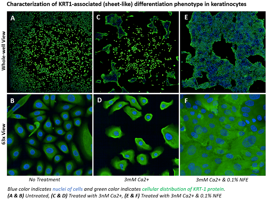
Study: To understand the effect of NFE on differentiating keratinocytes, we recreated the calcium-induced sheet-like structures6 and tracked the expression of KRT-1 protein. Using advanced cellular imaging technologies and image-classifier algorithms, we quantified the keratinocyte differentiation phenotype by virtue of the expression and morphological distribution of KRT-1 protein.
Observations: In the figure below, compared to untreated keratinocytes (A, B), exposure to calcium alone induced noticeable appearance of sheet-like structures and a moderate increase in KRT-1 protein expression (C, D), but when NFE was added to calcium-induced differentiating keratinocytes, we observed a significant enhancement of the sheet-like structures and an upregulation in KRT-1 protein expression (E, F). Since the distribution of KRT-1 protein closely followed the sheet-like structures, we called it the KRT1-associated differentiation phenotype.
Conclusions: These results suggest that NFE, by way of triggering enhanced KRT1-associated differentiation phenotype, could help in maintaining epithelial barrier function and skin integrity. Further investigation is required to substantiate NFE’s functional role.
References
- Autophagy activators stimulate the removal of advanced glycation end products in human keratinocytes.
JEADV 34 (Suppl. 3), 12-18 (2020); DOI: 10.1111/jvd.16453 - Chapter 1 – Skin Structure and Function.
Applied Dermatotoxicology. Clinical Aspects, 1-10 (2014); DOI: 10.1016/B978-0-12-420130-9 - Keratin 6, 16 and 17—Critical Barrier Alarmin Molecules in Skin Wounds and Psoriasis.
Cells 8, 807-821 (2019); DOI: 10.3390/cells8080807 - Keratin-1 maintains skin integrity and participates in an inflammatory network in skin through interleukin-18.
J Cell Sci. 125, 5269–5279 (2012); DOI: 10.1242/jcs.116574 - Calcium – a central regulator of keratinocyte differentiation in health and disease.
Eur J Dermatol 24, 650–661 (2014); DOI: 10.1684/ejd.2014.2452 - Monitoring calcium-induced epidermal differentiation in vitro using multiphoton microscopy.
J Biomed Opt. 25(7), 1-11 (2020); DOI: 10.1117/1.JBO.25.7.071205
The material and information contained on this webpage is for general information purposes only and subject to our Terms of Use.